

Spanish AI tool distinguishes Ewing sarcoma from similar tumors with 91.6% accuracy
A Spanish research team has developed and validated a deep-learning pathology tool that distinguishes Ewing sarcoma from morphologically similar tumors, with a reported overall accuracy of 91.6%. The practical selling point is not algorithmic novelty but tissue: the tool is intended to help pathologists choose the right confirmatory tests when the needle biopsy leaves very little tissue to work with.
What's new
A team from the Universitat Politècnica de València (UPV) and the University of Valencia has developed an artificial intelligence system that, according to the researchers, distinguishes Ewing sarcoma from other morphologically similar tumors with high accuracy. The model achieves 91.6% overall accuracy, 97.1% sensitivity specifically for Ewing sarcoma, and a marginal error rate of just 1.96% between Ewing sarcoma and rhabdomyosarcoma — the most difficult differential diagnosis among small round cell tumors, according to the team. The figures are given in UPV's press release and its republications on EurekAlert (source 155c25d2) and News-Medical (source 7ed56506).
The peer-reviewed article was published in the International Journal of Molecular Sciences on 31 July 2026 (Giner et al., volume 27, issue 15, article 6864, DOI 10.3390/ijms27156864). The institutions then distributed the press release in October 2026 — UPV on 1 October, EurekAlert on 8 October and News-Medical on 13 October — meaning the story is now live on the news cycle (source 9a1ba886).
Why the diagnosis is difficult
Ewing sarcoma belongs to the group of small round cell tumors, in which several tumor types look almost identical under the microscope. That means the pathologist can rarely make the diagnosis on morphology alone; it must be confirmed with molecular and other tests. Such confirmatory tests require tissue — and the sample is often taken by needle biopsy, which yields very limited tissue material.
This is where the tool has its practical entry point. "The tool is designed to support medical staff in the field of anatomical pathology and could help guide the tests needed to confirm the diagnosis, particularly when very little tissue is available from a biopsy. Using deep learning, the system identifies the microscopic patterns associated with each tumor type and then generates a classification," explains Valery Naranjo, coordinator of CVBLab at UPV and chief technology officer at the spin-off company Artikode Intelligence (source 155c25d2).
In practice, this means the classification can help the pathologist prioritize which of the limited tissue samples are used for which tests — an argument about "full optimisation of the tissue in biopsies" that UPV's release highlights, but which is the developer's own claim without prospective data in the available material.
How the system was built and tested
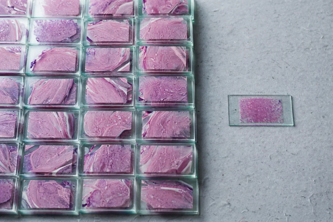
For training and evaluation, the team used 1,926 digitized histological samples from 729 patients at four hospitals in Spain and Italy. The material included 517 cases of Ewing sarcoma in addition to other tumor types (source 9a1ba886). The article title indicates that the analysis is based on tissue microarrays — standardized, small tissue samples assembled in a single preparation, which facilitates large-scale analysis but differs from whole slides from routine diagnostics.
The press release does not provide details on train/test splits, external validation, or which neighboring tumor types were included in the comparison beyond rhabdomyosarcoma as the hardest differential. Such validation details cannot be verified from the available material, since the article itself was not available for review here — only the press releases.
The researchers' own framing
Lead researcher and first author Francisco Giner (University of Valencia) pushes back on the idea that the news is the use of AI as such. "The novelty of the project does not lie simply in using AI to diagnose Ewing sarcoma, since experimental approaches based on deep learning already exist in this field. Our distinctive contribution lies in the combination of the clinical problem addressed, the type of data, the classification strategy and the focus on developing a tool that can be applied in the real-world setting of digital pathology," he stresses (source 7ed56506).
At the same time, he emphasizes the tool's role: artificial intelligence "is not intended to replace the pathologist, but rather to provide objective diagnostic support, which is particularly valuable in hospitals that do not have immediate access to the full range of molecular tests" (same source). That is a real health-policy point: molecular diagnostics require equipment and expertise that not all hospitals have, and morphological support from a deep-learning tool can therefore help where test capacity is limited.
Funding and partners
The work is part of the DEISA project (references INREIA/2024/71 and INREIA/2024/161), funded by the EU through NextGenerationEU funds. Partners are Hospital Universitari i Politècnic La Fe, the INCLIVA research institute, the Valencian Institute of Oncology (IVO), the Príncipe Felipe Research Center (CIPF), CIBERONC and the Rizzoli Orthopaedic Institute in Bologna (source 9a1ba886).
Caveats and open questions
Several things remain to be clarified before the figures can be read as a clinical statement:
- All performance figures come from a single press release. The UPV, EurekAlert and News-Medical documents are essentially the same release, not independent corroboration. The peer review applies to the article, not the press release's formulations of the figures.
- The validation details are unknown. The available material says nothing about train/test splits, external validation, or the definition of the comparator tumors — central questions for assessing whether 91.6% is a robust or optimistic value.
- Clinical status is unstated. The release describes a validated research system, but does not say whether the tool is in use, under regulatory review, or approved. There is no information supporting an assumption of clinical deployment.
- The timeline deserves precision. The article was published on 31 July 2026, but the press release only circulated in October 2026. That is not unusual, but it means the story's "news" on the news cycle is now the institutional rollout of a peer-reviewed result, not the publication itself.
- The claim of "full optimisation" of biopsy tissue is the developer's framing; there are no prospective data in the available material quantifying how much tissue the tool actually saves, or how often the classification changes test selection in practice.
This is, then, a credible, well-funded and peer-reviewed research result with a concrete clinical use case — but open questions about validation, regulation and actual clinical utility remain before the reported sensitivity of 97.1% can be translated into changed patient pathways.